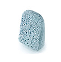
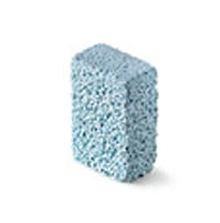
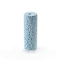
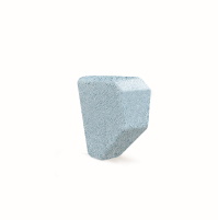
ENGIpore

sostituto osseo performato

ENGIpore-sostituto osseo performato

Elevata porosità

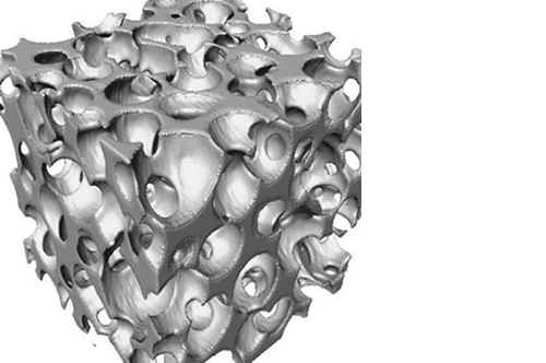
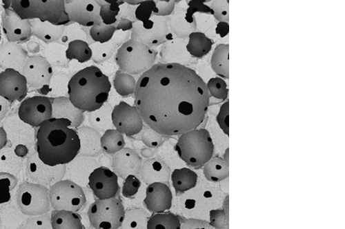
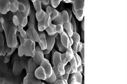
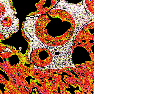
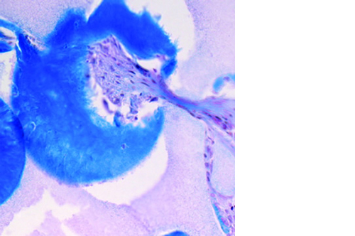
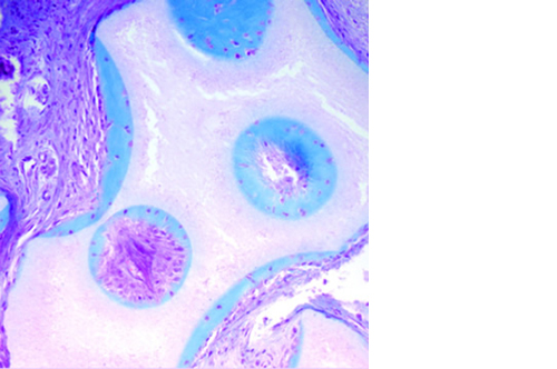
L’osteointegrazione del biomateriale e la neossificazione sono garantite da una struttura altamente porosa definita e controllata a livello macro, micro e di interconnessione dei pori.

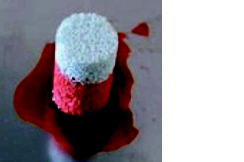
La porosità totale di ENGIpore può arrivare fino al 90%, favorendo un rapido assorbimento dei fluidi biologici e delle molecole segnale e permettendo la rapida colonizzazione cellulare indispensabile per una buona rigenerazione ossea.

Nonostante l’elevata porosità, ENGIpore è in grado di resistere a carichi in compressione al pari dell’osso spongioso.

Rapida osteointegrazione

Una volta applicato in situ, ENGIpore assorbe rapidamente le proteine bio-attive, i fattori di crescita e le cellule indifferenziate contenute nei liquidi fisiologici, elementi indispensabili per una rapida ed efficace rigenerazione ossea.

Applicazioni cliniche

ENGIpore è stato progettato per essere applicato in varie procedure chirurgiche:

021-indicazioni - indicazioni

ENGIpore può essere miscelato con osso autologo, fuidi biologici, concentrato midollare e fattori di crescita.

ENGIpore va inteso come un riempitivo di cavità o lacune ossee. Non deve sostituire le procedure standard per il trattamento dei difetti ossei. Deve essere utilizzato con gli opportuni mezzi di sintesi e stabilizzazione meccanica.

ENGIpore Ortho è disponibile con i seguenti codici:

Chips

codice
descrizione
PFS015005-40-00
1,0 - 2,5 mm - 5 cc
PFS015005-41-00
1,0 - 2,5 mm - 15 cc
PFS015005-50-00
2,5 - 4,0 mm - 5 cc
PFS015005-51-00
2,5 - 4,0 mm - 15 cc
PFS015005-52-00
2,5 - 4,0 mm - 30 cc

Cunei

codice
descrizione
PFS015005-30-00
40x30 mm, incl. 11° (h 10,5 mm)
PFS015005-31-00
40x30 mm, incl. 15° (h 13,5 mm)
PFS015005-32-00
40x30 mm, incl. 19° (h 16,5 mm)
PFS015006-19-00
30x15 mm, incl. 11° - 2 pcs. (h 8,5 mm)
PFS015006-20-00
30x15 mm, incl. 15° - 2 pcs. (h 10,5 mm)
PFS015006-25-00
30x15 mm, incl. 9° - 2 pcs. (h 7,5 mm)

Blocchetti

codice
descrizione
PFS015005-21-00
10x10x12 mm
PFS015005-22-00
20x10x12 mm
PFS015005-23-00
30x20x12 mm
PFS015005-24-00
40x20x12 mm

Cilindri cavi

codice
descrizione
PFS015008-01-00
ED = 12 mm ID = 3 mm h = 40 mm
PFS015008-02-00
ED = 15 mm ID = 3 mm h = 40 mm)
PFS015008-03-00
ED = 18 mm ID = 3 mm h = 40 mm)

SH

codice
descrizione
PFS015005-77-00
SH-Small 38 x 25 x 17 - 13 mm
PFS015005-76-00
SH-Medium 41 x 28 x 18 - 14 mm
PFS015005-75-00
SH-Large 45 x 30 x 20 - 15 mm

ENGIpore Spine è disponibile con i seguenti codici:

Chips

codice
descrizione
PFS015009-02-00
1,0 - 2,5 mm - 15 cc
PFS015009-03-00
2,5 - 4,0 mm - 5 cc
PFS015009-04-00
2,5 - 4,0 mm - 15 cc
  1. P.D. Parchi; M. Simonetti; E. Bonicoli; N. Piolanti; M. Scaglione: “Synthetic Bone Grafting in Aseptic Loosening of Acetabular Cup: Good Clinical and Radiological Outcomes in Contained Bone Defects at Medium-Term Follow up”. Int. J. Environ. Res. Public Health, 4;17(15):5624, 2020 
  2. Lovati, A. B. et al. In Vivo Bone Formation Within Engineered Hydroxyapatite Scaffolds in a Sheep Model. Calcified Tissue International 99, 209–223 (2016).
  3. Arrigoni, E. et al. Adipose-derived stem cells and rabbit bone regeneration: histomorphometric, immunohistochemical and mechanical characterization. Journal of Orthopaedic Science 18, 331–339 (2013).
  4. Manfrini, M. et al. Mesenchymal stem cells from patients to assay bone graft substitutes. Journal of Cellular Physiology 228, 1229–1237 (2013).
  5. Mastrangelo, F. et al. A Comparison of Bovine Bone and Hydroxyapatite Scaffolds During Initial Bone Regeneration: An In Vitro Evaluation. Implant Dentistry 22, 613–622 (2013).
  6. De Girolamo, L. et al. Role of autologous rabbit adipose-derived stem cells in the early phases of the repairing process of critical bone defects. Journal of Orthopaedic Research 29, 100–108 (2011).
  7. Güven, S. et al. Engineering of large osteogenic grafts with rapid engraftment capacity using mesenchymal and endothelial progenitors from human adipose tissue. Biomaterials 32, 5801–5809 (2011).
  8. Manfrini, M., et al. New generation of orthopaedic mimetic bioceramics assayed with human mesenchymal stem cells. European Musculoskeletal Review 6, (2011).
  9. Komlev, V. S. et al. Biodegradation of porous calcium phosphate scaffolds in an ectopic bone formation model studied by X-ray computed microtomograph. Eur Cell Mater 19, e46 (2010).
  10. Müller, A. M. et al. Towards an intraoperative engineering of osteogenic and vasculogenic grafts from the stromal vascular fraction of human adipose tissue. European cells & materials 19, 127–135 (2010).
  11. Tortelli, F., et al. The development of tissue-engineered bone of different origin through endochondral and intramembranous ossification following the implantation of mesenchymal stem cells and osteoblasts in a murine model. Biomaterials 31, 242–249 (2010).
  12. Manfrini, M., et al. High Porosity Bioceramic is a Favourable Environment for the Adhesion and Proliferation of Human Mesenchymal Stem Cells. INTERNATIONAL CONFERENCE ON TISSUE ENGINEERING, Leiria, Portugal, July 9-11, 2009
  13. Paderni, S., et al. Major bone defect treatment with an osteoconductive bone substitute. MUSCULOSKELETAL SURGERY 93, 89–96 (2009).
  14. Scheufler, O. et al. Spatial and temporal patterns of bone formation in ectopically pre-fabricated, autologous cell-based engineered bone flaps in rabbits. Journal of cellular and molecular medicine 12, 1238–1249 (2008).
  15. Marcacci, M. et al. Stem Cells Associated with Macroporous Bioceramics for Long Bone Repair: 6- to 7-Year Outcome of a Pilot Clinical Study. Tissue Engineering 13, 947–955 (2007).
  16. Timmins, N. E. et al. Three-Dimensional Cell Culture and Tissue Engineering in a T-CUP (Tissue Culture Under Perfusion). Tissue Engineering 13, 2021–2028 (2007).
  17. Scaglione, S. et al. Engineering of osteoinductive grafts by isolation and expansion of ovine bone marrow stromal cells directly on 3D ceramic scaffolds. Biotechnology and Bioengineering 93, 181–187 (2006).
  18. Mastrogiacomo, M. et al. Tissue engineering of bone: search for a better scaffold. Orthodontics & craniofacial research 8, 277–284 (2005).
  19. Marcacci M. et al. Repair of large bone defects with the use of autologous bone marrow stromal cells. N. Engl. J. Med. 344, 385–386 (2001).
  20. Kon, E. et al. Autologous bone marrow stromal cells loaded onto porous hydroxyapatite ceramic accelerate bone repair in critical-size defects of sheep long bones. Journal of biomedical materials research 49, 328–337 (2000).
  21. Chistolini, P. et al. Biomechanical evaluation of cell-loaded and cell-free hydroxyapatite implants for the reconstruction of segmental bone defects. J Mater Sci Mater Med 10, 739–742 (1999).
  22. Marcacci, M. et al. Reconstruction of extensive long-bone defects in sheep using porous hydroxyapatite sponges. Calcified tissue international 64, 83–90 (1999).
  23. Casabona, F. et al. Prefabricated engineered bone flaps: an experimental model of tissue reconstruction in plastic surgery. Plast. Reconstr. Surg. 101, 577–581 (1998).
  24. Muraglia, A., Martin, I., Cancedda, R. & Quarto, R. A Nude Mouse Model for Human Bone Formation in Unloaded Conditions. Bone 22, 131S–134S (1998).
  1. G. Barbanti Brodano; F. Lupidi; V. Tardivo; M. Bruzzo; A. Verlicchi; B. Zanotti: “Critical Evaluation of the Scientific Literature Concerning Bone Graft Alternatives in Spinal Surgery and Focus on Bioceramics”. Surg Technol Int. 2022 Jun 23;41:sti41/1582. doi: 10.52198/22.STI.41.NS1582. Online ahead of print.PMID: 35738571
  2. Parchi, P. D et al. Synthetic Bone Grafting in Aseptic Loosening of Acetabular Cup: Good Clinical and Radiological Outcomes in Contained Bone Defects at Medium-Term Follow Up. IJERPH (2020)
  3. Giorgi P, et al. (2015) Use of a novel porous hydroxyapatite bone graft substitute in postero-lateral and interbody spinal fusion: clinical and radiographic analysis at 4 year follow up
    Progress in Neuroscience, Vol. 3, N 1-4, 2015
  4. Barbanti Brodano G, et al. (2015) “A post-market surveillance analysis of the safety of hydroxyapatite-derived products as bone graft extenders or substitutes for spine fusion.”
    Eur Rev Med Pharmacol Sci 19(19):3548–3555
  5. Barbanera A, et al. (2013) “Potential applications of synthetic bioceramic bone graft substitute in spinal surgery.”
    Progress in Neuroscience 97–104
  6. Manfrini M, et al. (2013) “Mesenchymal stem cells from patients to assay bone graft substitutes.”
    J Cell Physiol 228(6):1229–1237
  7. Barbanti Brodano G, et al. (2012) “Human mesenchymal stem cells and biomaterials interaction: a promising synergy to improve spine fusion.”
    European Spine Journal 21(S1):3–9
  8. Komlev VS, et al. (2010) “Biodegradation of porous calcium phosphate scaffolds in an ectopic bone formation model studied by X-ray computed microtomograph.”
    Eur Cell Mater 19(136):e46
ENGIpore