MaioRegen

sostituto osteocondrale

MaioRegen-sostituto osteocondrale

Composizione e struttura

MaioRegen è una matrice multi-strato, prodotta grazie ad un processo esclusivo e brevettato. 


Il prodotto, composto da collagene e idrossiapatite arricchita in magnesio, mima il tessuto cartilagineo e osteo-condrale, sia nella composizione chimica che nella geometria micro- e nano-strutturale.


MaioRegen è disponibile in tre diverse configurazioni: MaioRegen Prime, MaioRegen Slim e MaioRegen Chondro+ rappresentano soluzioni specifiche per i diversi stadi della patologia pre-artrosica.


Biomimetismo: rigenerazione tissutale guidata

A) Il tessuto danneggiato è rimosso per creare un alloggiamento che ospiti l’impianto e per garantire un adeguato afflusso di sangue midollare

B) MaioRegen si adatta perfettamente alla lesione, ricostituendo la continuità anatomica

C) La struttura biomimetica porosa e la composizione chimica favoriscono la migrazione e l’adesione delle cellule multipotenti provenienti dal midollo

D) Le cellule proliferano, differenziano e sintetizzano nuova matrice in base allo strato del dispositivo che hanno colonizzato. Il progressivo riassorbimento e il rimodellamento mediato dalle cellule favoriscono una rigenerazione completa dei diversi tessuti cartilaginei e subcondrali

020-indicazioni - indicazioni

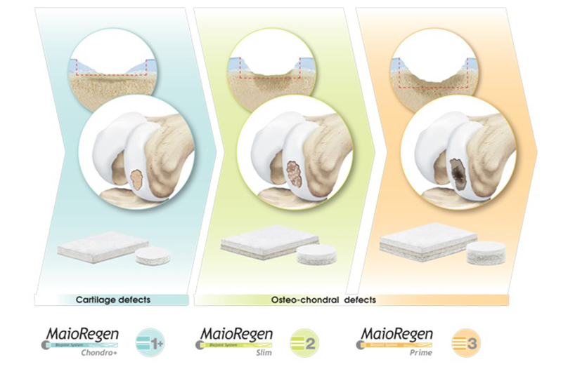
I dispositivi del Sistema MaioRegen sono indicati per il trattamento di lesioni articolari osteo-condrali e cartilaginee:

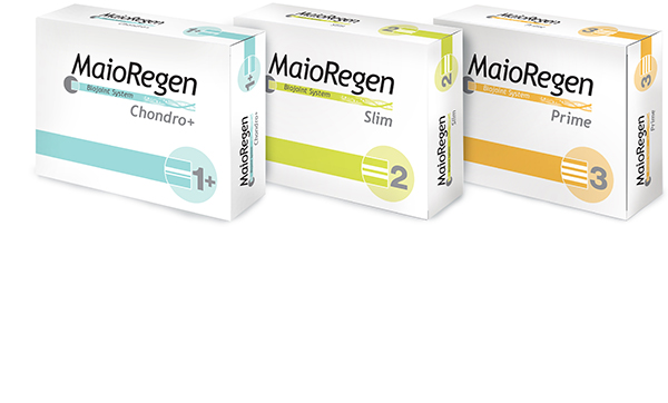
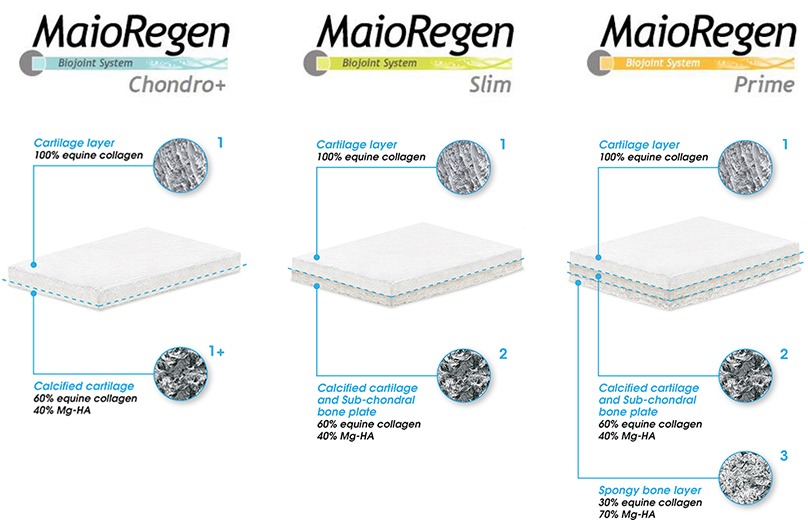
. MaioRegen Prime (scaffold tri-strato) è indicato per il trattamento di lesioni osteocondrali singole o multiple, con severa compromissione ossea, di grado IV secondo la classificazione di Outerbridge.
Le lesioni possono essere di origine traumatica, post-traumatica, degenerativa o derivanti da osteocondrite dissecante (OCD). 

 . MaioRegen Slim (scaffold bi-strato) è indicato per il trattamento di lesioni condrali profonde e osteocondrali, singole o multiple, con moderata compromissione ossea, di grado III e IV secondo la classificazione di Outerbridge. Le lesioni possono essere di origine traumatica, post-traumatica o degenerativa.

Entrambi i dispositivi sono altresì indicati per il trattamento di lesioni allo stadio di osteoartrosi precoce (Early OA), di grado I e II secondo la classificazione Kellgren e Lawrence, in assenza di osteofiti e in base al livello di compromissione ossea.

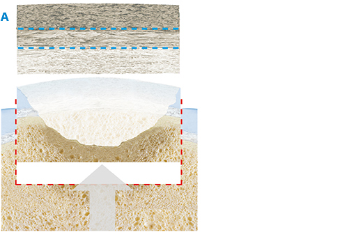
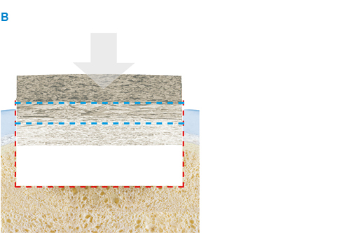
 . MaioRegen Chondro+ è indicato per il trattamento per il trattamento di lesioni condrali singole o multiple, con assente o lieve alterazione del tessuto osseo subcondrale, di grado III e IV secondo la classificazione di Outerbridge, di origine traumatica, post-traumatica o degenerativa.

MaioRegen Prime, MaioRegen Slim e MaioRegen Chondro+ sono forniti in confezioni singole e sterili, nei seguenti formati:

MaioRegen Chondro+

codice
descrizione
01600401
2,0x3,0x0,2 cm
1
01600400
3,0x4,0x0,2 cm
1
01600403
Ø = 1,2 cm H = 0,2 cm
1
01600405
Ø = 1,5 cm H = 0,2 cm
1
01600404
Ø = 1,8 cm H = 0,2 cm
1

MaioRegen Slim

codice
descrizione
01600289
2,0x3,0x0,4 cm
1
01600287
3,0x4,0x0,4 cm
1
01600290
Ø = 1,2 cm H = 0,4 cm
1
01600282
Ø = 1,5 cm H = 0,4 cm
1
01600292
Ø = 1,8 cm H = 0,4 cm
1

MaioRegen Prime

codice
descrizione
01600288
2,0x3,0x0,6 cm
1
01600286
3,0x4,0x0,6 cm
1
01600285
Ø = 1,2 cm H = 0,6 cm
1
01600275
Ø = 1,5 cm H = 0,6 cm
1
01600291
Ø = 1,8 cm H = 0,6 cm
1

MaioRegen Slim e MaioRegen Prime

Rimuovere il tessuto danneggiato e creare un alloggiamento regolare, con pareti parallele, mediate l’utilizzo di un osteotomo.

Creare un alloggiamento di una profondità adeguata: 4 - 5 mm per Maioregen Slim o 7 - 8 mm per Maioregen Prime.

Assicurarsi che il fondo dell’alloggiamento sia regolare.

Misurare accuratamente la dimensione della lesione.

Preparare il dispositivo in base alla dimensione della lesione precedentemente misura.

Utilizzare un bisturi per incidere lo strato superiore del dispositivo.

Utilizzare forbici chirurgiche per completare il taglio.

Si suggerisce l’applicazione di colla di fibrina sui bordi del dispositivo.

Nota bene: verificare l’orientamento del dispositivo identificando la faccia “ossea” caratterizzata da una superficie rugosa.

Posizionare il dispositivo assicurandosi che la superficie rugosa sia a contatto con l’osso, mentre la superficie liscia sia rivolta verso l’alto. Applicare una leggera pressione manuale.

Si suggerisce l’applicazione di colla di fibrina all’interfaccia superficiale dispositivo/cartilagine.

Verificare la stabilità del dispositivo mediate una serie di flesso/estensioni dell’articolazione.

MaioRegen Chondro+

Lodging Preparation 

Pulire e sagomare la lesione cartilaginea per creare un alloggiamento adatto all’impianto di MaioRegen Chondro+.

La profondità dell’alloggiamento per MaioRegen Chondro+ deve essere di 2.5 mm.

Procedere con un’appropriata tecnica di stimolazione midollare (es. microfratture, nanofratture, perforazioni) al fine di facilitare il sanguinamento dell’osso subcondrale.

Misurare accuratamente la dimensione dell’alloggiamento.

Preparare il dispositivo in base alla dimensione della lesione precedentemente misura.

Tagliare il dispositivo mediate utilizzo di una forbice chirurgica.

Posizionare il dispositivo nell’alloggiamento mediate una leggera pressione manuale, facendo particolare attenzione all’orientamento (superficie rugosa verso la parte ossea).

Si suggerisce l’applicazione di colla di fibrina all’interfaccia superficiale dispositivo/cartilagine.

Verificare la stabilità del dispositivo mediate una serie di flesso/estensioni dell’articolazione.

Ad oggi oltre 4.000 pazienti sono stati trattati con MaioRegen, che risulta essere un trattamento sicuro ed efficace.

Uno studio clinico randomizzato e controllato (vs. le microfratture) con Livello di Evidenza 1, dimostra un miglioramento statisticamente significativo negli score clinici (IKDC soggettivo, Tegner, KOOS, VAS) tra pre- e post-op (2 anni follow-up).

MaioRegen è risultato essere superiore alle microfratture per le seguenti indicazioni cliniche: lesioni osteocondrali profonde, pazienti attivi sportivamente e lesioni da osteocondrite dissecante (OCD).


Lesioni osteocondrali profonde

Superiorità statisticamente significativa di MaioRegen vs. Microfratture (+12.4 punti nel change from baseline per lo score IKDC soggettivo).


Osteocondrite Dissecante (OCD)

Superiorità di 12 punti nel change from baseline di MaioRegen vs. Microfratture per lo score IKDC soggettivo.


Pazienti sportivamente attivi

Superiorità statisticamente significativa di MaioRegen vs. Microfratture (+16 punti nel change from baseline per lo score IKDC soggettivo).

La superiorità in queste specifiche popolazioni fornisce indicazioni cliniche chiare per il trattamento con MaioRegen Prime.

Inoltre, uno studio pilota con 27 pazienti dimostra la stabilità degli score clinici a 8 anni dal trattamento con MaioRegen nel caso di pazienti con lesioni osteocondrali complesse.

IKDC Soggetivo – 96 mesi post-operatorio

Tegner Score – 96 mesi post-operatorio

Dati in-vitro e pre-clinici

  1. Vukasovic A.  et Al:  Bioreactor‐manufactured cartilage grafts repair acute and chronic osteochondral defects in large animal studies. Cell Proliferation- Wiley (2019).
  2. Calabrese G. et al. Combination of Collagen-Based Scaffold and Bioactive Factors Induces Adipose-Derived Mesenchymal Stem Cells Chondrogenic Differentiation In vitro. Front Physiol (2017).
  3. Calabrese G. et al. Collagen-Hydroxyapatite Scaffolds Induce Human Adipose Derived Stem Cells Osteogenic Differentiation In Vitro. PLoS One (2016). 
  4. Grigolo B. et al. Novel nano-composite biomimetic biomaterial allows chondrogenic and osteogenic differentiation of bone marrow concentrate derived cells. J Mater Sci Mater Med (2015).
  5. Tampieri A. et al. Mimicking natural bio-mineralization processes: a new tool for osteochondral scaffold development. Trends Biotechnol (2011).
  6. Kon E. et al. Novel nanostructured scaffold for osteochondral regeneration: pilot study in horses. J Tissue Eng Regen Med (2010).
  7. Kon E. et al. Orderly osteochondral regeneration in a sheep model using a novel nano-composite multilayered biomaterial. J Orthop Res (2010).
  8. Kon E. et al. Platelet autologous growth factors decrease the osteochondral regeneration capability of a collagen-hydroxyapatite scaffold in a sheep model. BMC Musculoskeletal Disorders (2010).
  9. Tampieri A. et al. Design of graded biomimetic osteochondral composite scaffolds. Biomaterials (2008).
  10. Tampieri A. et al. Biomimetic Hybrid Composites to Repair Osteochondral Lesions. KEM (2008).

Letteratura clinica

  1. Ricci M. et al. Cell-free osteochondral scaffolds provide a substantial clinical benefit in the treatment of osteochondral defects at a minimum follow-up of 5 years. J EXP ORTOP (2021).
  2. Di Martino A. et al. Cell-Free Biomimetic Osteochondral Scaffold for the Treatment of Knee Lesions: Clinical and Imaging Results at 10-Year Follow-up. Am J Sports Med (2021). 
  3. Casiraghi A. et al. A case report of femoral head fracture with osteochondral lesion treated by osteosynthesis and biomimetic scaffold: 2-year clinical and radiological follow-up. J EXP ORTOP (2021). 
  4. Botond G. et al. Three-dimensional acellular scaffolds (MaioRegen) in isolated chondral defect. The publishing house medicine of the romanian academy (2021). 
  5. Schreiner M. M. et al. Reliability of the MOCART (Magnetic Resonance Observation of Cartilage Repair Tissue) 2.0 knee score for different cartilage repair techniques—a retrospective observational study. Eur Radiol (2021). 
  6. Sessa A. et Al:  Treatment of Juvenile Knee Osteochondritis Dissecans with a Cell-Free Biomimetic Osteochondral Scaffold: Clinical andMRI Results at Mid-Term Follow-u.  Cartilage (2020).
  7. Martincic D. et Al: Autologous chondrocytes versus filtered bone marrow mesenchymal stem/stromal cells for knee cartilage repair—a prospective study. Int_Ortho (2020).
  8. Veber M. et Al: Combination of Filtered Bone Marrow Aspirate and Biomimetic Scaffold for the Treatment of Knee Osteochondral Lesions: Cellular and Early Clinical Results of a Single Centre Case Series. Tissue _Eng_Regen Med (2020).
  9. Guerin g et Al.: Repair of large condylar osteochondral defects of the knee by collagenscaffold. Minimum two-year outcomes. Ortho. & Trauma. (2020).
  10. Sessa A. et Al: Cell-Free Biomimetic Osteochondral Scaffold Implantation Technique. JBJS (2019).
  11. Sessa A. et Al.: Cell-Free Osteochondral Scaold for the Treatment of Focal Articular Cartilage Defects in Early Knee OA:  5 Years’ Follow-Up Results. JCM (2019).
  12. Mrtincic D et Al.:  Survival Rates of Various Autologous Chondrocyte Grafts and Concomitant Procedures.  Prospective Single-Center  Study over 18 Years.  Cell transplantation (2019).
  13. D’ambrosi R.  et Al.: MaioRegen Osteochondral Substitute for the Treatment of Knee Defects: A systematic Review of the Literature. JCM (2019).
  14. Gabusi E. et Al.: Cartilage and Bone Serum Biomarkers as Novel Tools for Monitoring Knee steochondritis Dissecans Treated with Osteochondral Scaffold. BRI (2018).
  15. Condello V. et Al.: Use of a Biomimetic Scaffold for the Treatment of Osteochondral Lesions in Early Osteoarthritis. BMRI (2018).
  16. Kaipel M. et Al.: Beneficial clinical effects but limited tissue quality following osteochondral repair with a cell-free multi-layered nano-composite scaffold in the talus. FAS (2016).
  17. Perdisa F. et Al.: Treatment of Knee Osteochondritis Dissecans With a Cell-Free Biomimetic Osteochondral Scaffold. AJSM (2017).
  18. Mathis D. et Al.: Good clinical results but moderate osseointegration and defect filling of a cell‑free multi‑layered nano‑composite scaffold for treatment of osteochondral lesions of the knee. KSSTA (2017).
  19. Albano D. et Al.: Clinical and imaging outcome of osteochondral lesions of the talus treated
    using autologous matrix-induced chondrogenesis technique with a biomimetic scaffold. BMCMD (2017).

  20. Kon E. et al. A multilayer biomaterial for osteochondral regeneration shows superiority vs microfractures for the treatment of osteochondral lesions in a multicentre randomized trial at 2 years. Knee Surg Sports Traumatol Arthrosc (2017).

  21. Perdisa F. et al. One-Step treatment for patellar cartilage defects with a cell-free osteochondral scaffold. A prospective clinical and MRI evaluation. Am J Sports Med (2017).
  22. Berruto M. et al. Can a biomimetic osteochondral scaffold be a reliable alternative to prosthetic surgery in treating late-stage SPONK? Knee (2016).
  23. Brix M. et al. Successful osteoconduction but limited cartilage tissue quality following osteochondral repair by a cell-free multilayered nano-composite scaffold at the knee. Int Orthop (2016).
  24. Di Martino A. et al. Surgical treatment of early knee osteoarthritis with a cell-free osteochondral scaffold: results at 24 months of follow-up. Injury (2015).
  25. Verdonk P. et al. Treatment of Osteochondral Lesions in the Knee with a Cell-Free Scaffold. Bone Joint J (2015).
  26. Christensen BB. et al. Poor osteochondral repair by a biomimetic collagen scaffold: 1- to 3-year clinical and radiological follow-up. Knee Surg Sports Traumatol Arthrosc (2015).
  27. Persiani P. et al. Osteochondritis dissecans of the lateral femoral condyle in a patient affected by osteogenesis imperfecta: a case report. J Pediatric Orthop (2015).
  28. Delcogliano M. et al. Treatment of osteochondritis dissecans of the knee with a biomimetic scaffold. A prospective multicenter study. Joints (2014).
  29. Kon E. et al. Tibial plateau lesions. Surface reconstruction with a biomimetic osteochondral scaffold: Results at 2 years of follow-up. Injury (2014).
  30. Berruto M. et al. Treatment of Large Knee Osteochondral Lesions With a Biomimetic Scaffold: Results of a Multicenter Study of 49 Patients at 2-Year Follow-up. Am J Sports Med (2014).
  31. Kon E. et al. A one-step treatment for chondral and osteochondral knee defects: clinical results of a biomimetic scaffold implantation at 2 years of follow-up. J Mater Sci Mater Med (2014).
  32. Filardo G. et al. Fibrin glue improves osteochondral scaffold fixation: study on the human cadaveric knee exposed to continuous passive motion. Osteoarthr Cartil (2014).
  33. Filardo G. et al. Osteochondral scaffold reconstruction for complex knee lesions: a comparative evaluation. Knee (2013).
  34. Delcogliano M. et al. Use of innovative biomimetic scaffold in the treatment for large osteochondral lesions of the knee. Knee Surg Sports Traumatol Arthrosc (2013).
  35. Kon E. et al. Clinical Results and MRI Evolution of a Nano-Composite Multilayered Biomaterial for Osteochondral Regeneration at 5 Years. Am J Sports Med (2013).
  36. Filardo G. et al. Treatment of Knee Osteochondritis Dissecans With a Cell-Free Biomimetic Osteochondral Scaffold: Clinical and Imaging Evaluation at 2-Year Follow-up. Am J Sports Med (2013).
  37. Marcacci M. et al. Unicompartmental osteoarthritis: an integrated biomechanical and biological approach as alternative to metal resurfacing. Knee Surg Sports Traumatol Arthrosc (2013).
  38. Filardo G. et al. Midterm results of a combined biological and mechanical approach for the treatment of a complex knee lesion. Cartilage (2012).
  39. Kon E. et al. How to Treat Osteochondritis Dissecans of the Knee: Surgical Techniques and New Trends: AAOS Exhibit Selection. J Bone Joint Surg Am (2012).
  40. Kon E. et al. Novel Nano-composite Multilayered Biomaterial for Osteochondral Regeneration: A Pilot Clinical Trial. Am J Sports Med (2011).
  41. Kon E. et al. A novel nano-composite multi-layered biomaterial for treatment of osteochondral lesions: technique note and an early stability pilot clinical trial. Injury (2010).
  42. Kon E. et al. Novel nano-composite multi-layered biomaterial for the treatment of multifocal degenerative cartilage lesions. Knee Surg Sports Traumatol Arthrosc (2009).

Altre citazioni

  1. Ambra LF et al. Interventions for cartilage disease - current state of the art and emerging technologies. Arthritis Rheumatol (2017).
  2. Pina S. et al. Pre-clinical and Clinical Management of Osteochondral Lesions. Regenerative Strategies for the Treatment of Knee Joint Disabilities (Chapter 8, 2017).
  3. Brittberg M. et al. Cartilage repair in the degenerative ageing knee. Acta Orthopaedica (2016).
  4. Makhni EC. et al. Comprehensiveness of Outcome Reporting in Studies of Articular Cartilage Defects of the Knee. Arthroscopy (2016).
  5. Cross LM. et al. Nanoengineered biomaterials for repair and regeneration of orthopedic tissue interfaces. Acta Biomater (2016).
  6. Fischer S. et al. Single-step scaffold-based cartilage repair in the knee: A systematic review. J Orthop (2016).
  7. Jeuken RM. et al. Polymers in Cartilage Defect Repair of the Knee: Current Status and Future Prospects. Polymers (2016).
  8. Gobbi A. et al. Scaffolding as Treatment for Osteochondral Defects in the Ankle. Arthroscopy (2016).
  9. Vinatier C. et al. Cartilage tissue engineering: From biomaterials and stem cells to osteoarthritis treatments. Ann Phys Rehabil Med (2016).
  10. Angele P. et al. Chondral and osteochondral operative treatment in early osteoarthritis. Knee Surg Sports Traumatol Arthrosc (2016).
  11. Xuezhou L. et al. Biomimetic biphasic scaffolds for osteochondral defect repair. Regenerative Biomaterials (2015).
  12. Gobbi A. et al. Fresh osteochondral allografts in the knee: only savage procedure? Ann Transl med (2015).
  13. Di Luca A. et al. The osteochondral interface as a gradient tissue: from development to the fabrication of gradient scaffolds for regenerative medicine. Birth Defects Res (2015).
  14. Smith BD. et al. The current state of scaffolds for musculoskeletal regenerative applications. Nat Rev Rheumatol (2015).
  15. Wylie JD. et al. What Is the Effect of Matrices on Cartilage Repair? A Systematic Review. Clin Orthop Related Res (2015).
  16. Wylie JD. et al. Failures and Reoperations After Matrix-Assisted Cartilage Repair of the Knee: A Systematic Review. Arthroscopy (2015).
  17. Zanon G. et al. Osteochondritis dissecans of the knee. Joints (2014).
  18. Luthringer BJ. et al. Magnesium-based implants: a mini-review. Magnesium Research (2014).
  19. Yan LP. et al. Bilayered silk/silk-nanoCaP scaffolds for osteochondral tissue engineering: In vitro and in vivo assessment of biological performance. Acta Biomaterialia (2014).
  20. Atesok M. et al. Multilayer scaffolds in orthopaedic tissue engineering. Knee Surg Sports Traumatol Arthrosc (2014).
  21. Kon E. et al. Acellular Matrix-Based Cartilage Regeneration Techniques for Osteochondral Repair. Oper Techniques Orthop (2014).
  22. Kon E. et al. Clinical results of multilayered biomaterials for osteochondral regeneration. Journal of Experimental Orthopaedics (2014).
  23. Lee EJ. et al. Biomaterials for Tissue Engineering. Ann Biomed Eng (2014).
  24. Filardo G. et al. Scaffold-based repair for cartilage healing: a systematic review and technical note. Arthroscopy (2013).
  25. Kon E. et al. New trends for knee cartilage regeneration: from cell-free scaffolds to mesenchymal stem cells. Curr Rev Musculoskelet Med (2012).
  26. Kon E. et al. Current and future scaffolds for articular cartilage repair. Journal of Orthopedics (2012).
  27. Luyten FP. et al. Tissue engineering approaches for osteoarthritis. Bone (2012).
  28. Gomoll AH. et al. Surgical treatment for early osteoarthritis. Part I: cartilage repair procedures. Knee Surg Sports Traumatol Arthrosc (2011).
  29. Panseri S. et al. Osteochondral tissue engineering approaches for articular cartilage and subchondral bone regeneration. Knee Surg Sports Traumatol Arthrosc (2011).
  30. Gomoll AH. et al. The subchondral bone in articular cartilage repair: current problems in the surgical management. Knee Surg Sports Traumatol Arthrosc (2010).
Mini-arthrotomic surgical approach
Management options for OCL in the knee
Novel Biomimetic 3D Scaffold in the Knee
Management of Early Arthritis on the ankle
Regeneration Clinical results of a novel 3D Matrix
OCD treatment with MaioRegen
MaioRegen 3D animation
Dottori in prima linea
Case history. Poter tornare a ballare