Sintlife

mouldable bone substitute

Sintlife-mouldable bone substitute

Mg-substituted apatite: just as nature designed it

It has been demonstrated that the presence of Mg2+ enables the hydroxyapatite crystal cell structure to become unstable and biologically active, promoting rapid cell-mediated material resorption, bone formation and remodelling.

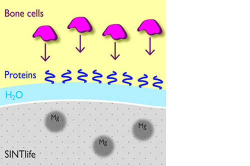
In addition, Mg2+ substituted apatite displays improved surface properties: the biomaterial effectively interacts with water molecules* to rapidly capture the key proteins involved in osteogenesis.

*Bertinetti et al. Langmuir (2009)


Synchronized process: material resorption and new bone formation

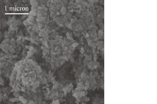
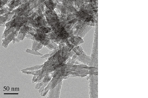
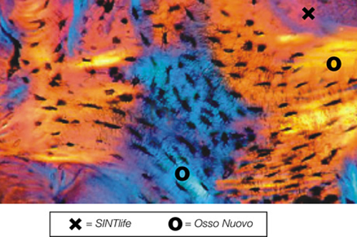
SINTlife is a unique material that interacts with bone-forming cells and promotes deposition of new bone tissue. Thanks to its specific biomimetic chemical composition, nano-structure and surface properties, SINTlife is remodelled and resorbed by cellular action over a period of 6-18 months, allowing for sufficient biostimulative scaffold to remain during the formation and maturation of new bone.

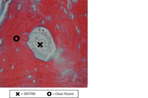
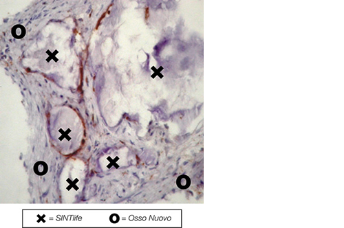
During the remodelling phase, osteoclastic resorption and osteoblastic osteogenenetic activities are seen around the remaining SINTlife particles*, up to a complete bone regeneration. As a result, SINTlife leads to a physiological, rapid and effective repair.

*Landi et al., J. Mater Sci: Mater Med (2008)

Clinical applications

SINTlife is designed for use in a broad range of procedures, such as:

021-indicazioni - indications

SINTlife may be combined with autologous bone, blood, bone marrow or growth factors.

SINTlife is not intended to modify or replace standard procedures for the treatment of bone defects, but for filling bony voids or gaps of the skeletal system, that are not intrinsic to the stability of the bony structure. It must be used with appropriate stabilising hardware.

SINTlife spine is available with the following codes:

Putty

code
description
PFS015057-08-00
Putty (1,5 cc syringe)
PFS015057-04-00
Putty (2,5 cc syringe)
PFS015057-05-00
Putty (5 cc syringe)

SINTlife Ortho is available with the following codes:

Putty

code
description
PFS015056-01-00
Putty (1 cc syringe)
PFS015056-02-00
Putty (2,5 cc syringe)
PFS015056-00-00
Putty (5 cc syringe)
PFS015056-05-00
Putty (10 cc syringe)
  1. Sartori M, et al. (2014) “Long-term in vivo experimental investigations on magnesium doped hydroxyapatite bone substitutes”.
    Journal of Materials Science: Materials in Medicine 25(6):1495–1504
  2. Dallari D, et al. (2012) “A prospective, randomised, controlled trial using a Mg-hydroxyapatite - demineralized bone matrix nanocomposite in tibial osteotomy”.
    Biomaterials 33(1):72–79
  3. Manfrini M, et al. (2011) “New Generation of Orthopaedic Mimetic Bioceramics Assayed with Human Mesenchymal Stem Cells”.
  4. Manfrini M, et al. (2009) “High porosity bioceramic is a favourable environment for the adhesion and proliferation of human mesenchymal stem cells”.
    ECCOMAS – INTERNATIONAL CONFERENCE ON TISSUE ENGINEERING 2009 P.J. Bártolo et al. (Eds.) Leiria, Portugal, July 9-11, 2009
  5. Landi E, et al. (2008) “Biomimetic Mg-substituted hydroxyapatite: from synthesis to in vivo behaviour”.
    Journal of Materials Science: Materials in Medicine 19(1):239–247
  1. B. Zanotti; F. Muggiolu; L. De Maria: “The benefit of antibiotic-combined Mg-hydroxyapatite bone graft substitute over autologous bone for surgical site infection prevention in posterolateral spinal fusion: a retrospective cohort study.” Annals of Medicine Surgery, 27;85(6):2341-2347, 2023
  2. C. Griffoni; V. Canella; G. Tedesco; A. Nataloni; A. Zerbi; A. Gasbarrini; G. Barbanti Brodano: “Ceramic bone graft substitute (Mg-HA) in spinal fusion: a prospective pilot study.” DOI 10.3389/fbioe.2022.1050495, Frontiers in Bioengineering and Biotechnology, 2022
  3. V. Cioffi; A. Bocchetti; A. Nataloni; V. Canella; M. Sandri; R. De Falco: “Anterior cervical fusion using magnesium-enriched hydroxyapatite: a two-year follow-up in 75 cases”. Vol. 6, 37-42, N. 1-4, Progress in Neuroscience, 2021
  4. B. Zanotti; F. Muggiolu; A. Nataloni; V. Canella: “Efficacy and safety of the magnesium-hydroxyapatite bone graft substitute in postero-lateral spinal fusion: observational, spontaneous clinical study”. Progress in Neuroscience, Vol. 6, 27-35, N. 1-4, 2021
  5. G. Barbanti Brodano; C. Griffoni; B. Zanotti; A. Gasbarrini; S. Bandiera; R. Ghermandi, S. Boriani: “A Post-Market Surveillance Analysis of the safety of hydroxyapatite-derived products as bone graft extenders or substitutes for spine fusion”. European Review for Medical and Pharmacological Sciences, 2015; 19:3548-3555 
  6. Barbanti Brodano G, et al. (2015) “A post-market surveillance analysis of the safety of hydroxyapatite-derived products as bone graft extenders or substitutes for spine fusion.” 
    Eur Rev Med Pharmacol Sci 19(19):3548–3555
  7. Barbanti Brodano G, et al. (2014) “Hydroxyapatite-Based Biomaterials Versus Autologous Bone Graft in Spinal Fusion: An In Vivo Animal Study.” 
    Spine 39(11):E661–E668
  8. Barbanera A, et al. (2013) “Potential applications of synthetic bioceramic bone graft substitute in spinal surgery.” 
    Progress in Neuroscience 97–104
  9. Manfrini M, Di et al. (2013) “Mesenchymal stem cells from patients to assay bone graft substitutes.” 
    Journal of Cellular Physiology 228(6):1229–1237
  10. Barbanti Brodano G, et al. (2012) “Human mesenchymal stem cells and biomaterials interaction: a promising synergy to improve spine fusion.” 
    European Spine Journal 21(S1):3–9
  11. Pola E, et al. (2011) “Bioplasty for vertebral fractures: preliminary results of a pre-clinical study on goats using autologous modified skin fibroblasts.” 
    International journal of immunopathology and pharmacology 24(1 Suppl 2):139
Sintlife