Regenoss

flexible bone substitute

Regenoss-flexible bone substitute

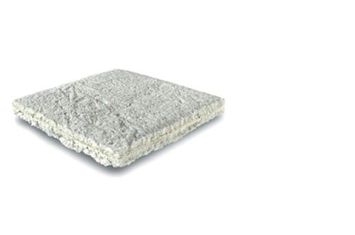
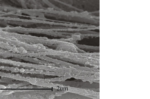
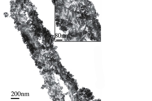
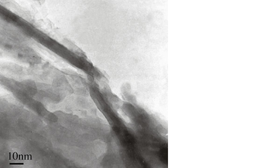
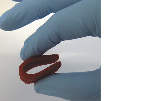
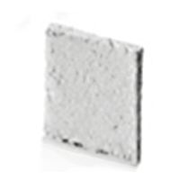
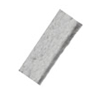
Biomimetic nano-structure: as modelled in nature

RegenOss is a fully biomimetic scaffold, its unique structure and chemical composition confer characteristics replicating that of human bone.

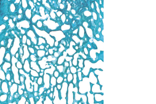
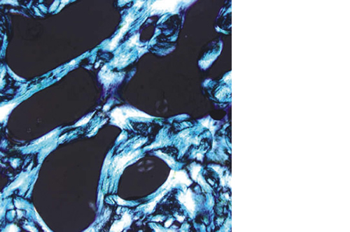
By using nature as a model, the patented nucleation of magnesium-enriched hydroxyapatite nano-crystals into type I collagen fibers resembles the process occurring during biological neo-ossification.

Replaced by new bone

RegenOss is highly hydrophilic; it can quickly absorb the biological fluids, molecules and cells promoting bone formation. Its architecture favors cells attachment and proliferation.

While serving as a scaffold to guide effective bone regeneration, RegenOss is resorbed by enzymes and cellular action over a period of 6-12 months.

Clinical applications

RegenOss is designed for use in a broad range of procedures, such as:

021-indicazioni - indications

RegenOss may be combined with autologous bone, blood, bone marrow or growth factors.

RegenOss is not intended to modify or replace standard procedures for the treatment of bone defects, but for filling bony voids or gaps of the skeletal system, that are not intrinsic to the stability of the bony structure. RegenOss must be used with appropriate stabilising hardware.

RegenOss Ortho is available with the following codes: 

Patch

code
description
01600816
3,5 x 3,5 x 0,5 cm
01600818
2,5 x 7,5 x 0,5 cm
01600824
6,5 x 6,5 x 0,5 cm

RegenOss Spine is available with the following codes: 

Strip

code
description
01600821
2,5 x 7,5 x 0,5 cm
  1. P. Cimatti, I. Andreoli, M. Busacca, M. Govoni, L. Vivarelli, N. Del Piccolo, A. Maso, C. Stagni, G. Pignatti: “An Observational Prospective Clinical Study for the Evaluation of a Collagen-Hydroxyapatite Composite Scaffold in Hip Revision Surgery” J. Clin. Med. 2022, 11, 6372. https://doi.org/10.3390/jcm11216372. Free PMC article.  
  2. Romano' CL, et al. (2015) “Antibiotic-loaded RegenOss for the treatment of septic bone defects in vitro study and preclinical experience”
    JOURNAL OF BIOLOGICAL REGULATORS & HOMEOSTATIC AGENTS Vol. 29, no. 3 (S), 0-0 (2015)
  3. Babiker, H., Ding, et al. (2012) “The effects of bone marrow aspirate, bone graft, and collagen composites on fixation of titanium implants.”
    J. Biomed. Mater. Res. Part B Appl. Biomater. 100, 759–766 (2012).
  4. Arrigoni E, et al. (2013) “Two bone substitutes analyzed in vitro by porcine and human Adipose-derived Stromal Cells”
    Int J Immunopathol Pharmacol. 2013 Jan-Mar;26(1 Suppl):51-9.
  1. P. Cimatti, I. Andreoli, M. Busacca, M. Govoni, L. Vivarelli, N. Del Piccolo, A. Maso, C. Stagni, G. Pignatti: “An Observational Prospective Clinical Study for the Evaluation of a Collagen-Hydroxyapatite Composite Scaffold in Hip Revision Surgery” J. Clin. Med. 2022, 11, 6372. https://doi.org/10.3390/jcm11216372. Free PMC article. (ORTHO)
  2. G. Gioia, M. Agnoletto, A. Di Giancamillo, M. Domenicucci, L. Mangiavini, M.D.M. Lombrardo, L. Brambilla, G.M. Peretti: “Histological assessment of new bone formation with biomimetic scaffold in posterolateral lumbar spine”. J Biol Regul Homeost Agents; Jul-Aug 2020; 34(4 Suppl. 3):99-103, 2020 (SPINE)
  3. A. Barbanera, M. Vitali, A. Nataloni, V. Canella: “Clinical performance of a collagen-based hydroxyapatite bone graft substitute in procedures of spinal arthrodesis”. Progress in Neuroscience; 1 (1-4): 117-124, 2020 (SPINE) 
  4. P. Giorgi; D. Capitani; S. Sprio; M. Sandri; A. Tampieri; V. Canella; A. Nataloni; G. Schirò: “A new bioinspired collagen-hydroxyapatite bone graft substitute in adult scoliosis surgery:  results at 3 years follow-up”. Journal App. Biomaterial 2017 (SPINE)
  5. B. Grigolo, P. Dolzani, M. Tenucci, C. Giannetti, G. Calvosa: “Histologically demonstrated posterolateral spine fusion using a collagen-hydroxyapatite bone substitute in human: a case report.” International Journal of Clinical and Experimental Medicine, 2016
  6. Barbanti Brodano G, et al. (2015) “A post-market surveillance analysis of the safety of hydroxyapatite-derived products as bone graft extenders or substitutes for spine fusion.”
    Eur Rev Med Pharmacol Sci 19(19):3548–3555
  7. Barbanera A, et al. (2013) “Potential applications of synthetic bioceramic bone graft substitute in spinal surgery.”
    Progress in Neuroscience 97–104
  8. Manfrini M, et al. (2013) “Mesenchymal stem cells from patients to assay bone graft substitutes.”
    J Cell Physiol 228(6):1229–1237
  9. Grigolo B. et al. (2016) “Use of a fully-resorbable, biomimetic composite hydroxyapatite as bone graft substitute for posterolateral spine fusion: a case report”.
    Int J Clin Exp Med 2016;9(11):22458-22462
Regenoss