MaioRegen

osteochondral substitute

MaioRegen-osteochondral substitute

Composition and structure

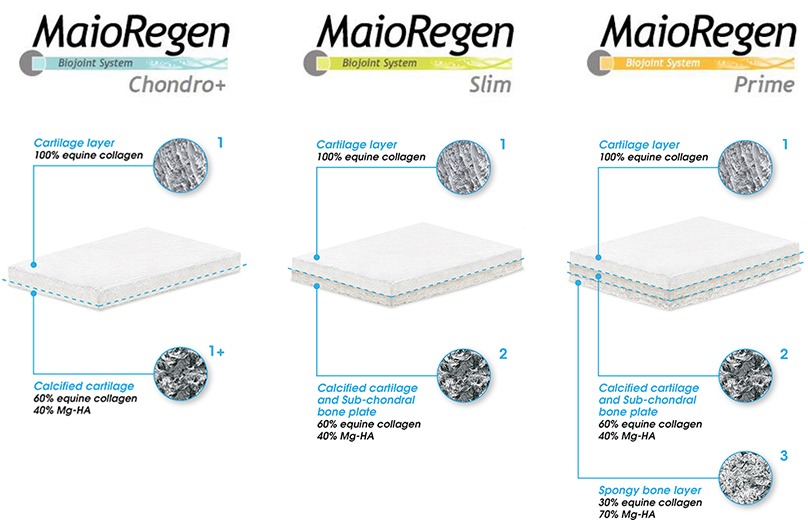
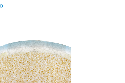
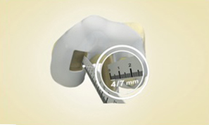
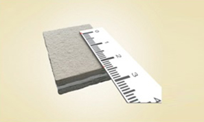
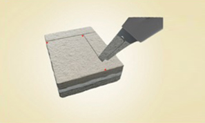
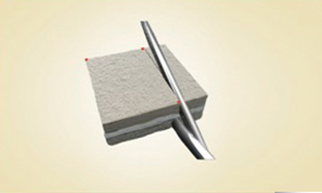
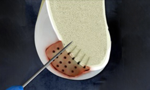
MaioRegen is a multi-layered matrix, manufactured through a patented process.

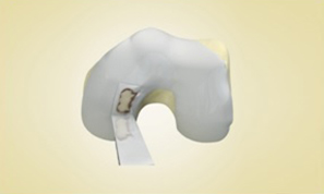
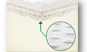
The product, composed by collagen and hydroxyapatite enriched with magnesium, mimics the chondral and osteochondral tissues, both in the chemical composition and in the micro- and nano-structure.

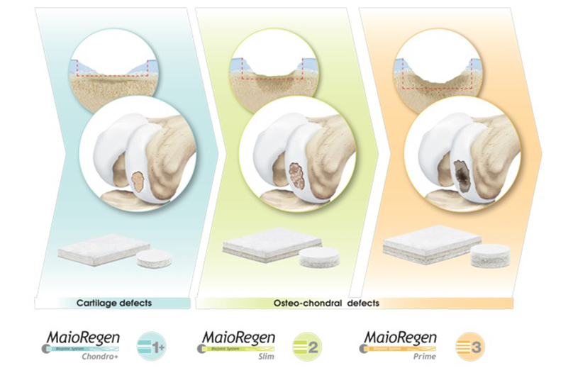
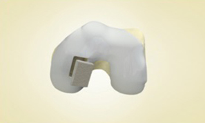
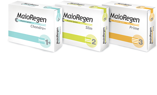
MaioRegen is available in three different configurations: MaioRegen Prime, MaioRegen Slim and MaioRegen Chondro+ represent specific solutions for the treatment of the different phases of early phases of arthritic pathology.


Biomimetism

Cartilage layer

Calcified cartilage and sub-chondral bone layer

Spongy bone layer

A) The damaged tissue is removed to create a suitable lodging to host the implant and guarantee an adequate blood flow from the subchondral bone

B) Maioregen fits perfectly into lesion site, restoring anatomic continuity

C) The biomimetic porous structure and the chimical composition favour cellular migration. The bone marrow-derived cells selectively adhere to the collagen fibres, colonizing the entire scaffold

D) Cells proliferate, differentiate and synthesize matrix according to scaffold layer composition. The progressive resorption of MaioRegen and the simultaneous cell mediated remodeling favour the complete regeneration of the tissue

020-indicazioni - indications

The devices of MaioRegen system are indicated for the treatment of cartilage and osteochondral joint surface lesions

 . MaioRegen Prime (tri-layer) is indicated for the treatment of single/multiple osteochondral lesions with severely compromised bone, Outerbridge Grade IV. Lesions can be f traumatic, post-traumatic or degenerative origin, as well as caused by osteochondritis dissecans (OCD).

 . MaioRegen Slim (bi-layer) is indicated for the treatment of single/multiple osteochondral lesions with slightly compromised bone, Outerbridge Grade III and IV. Lesions can be f traumatic, post-traumatic or degenerative origin.

Both devices are also indicated for Early OA lesions, Grade I/II according to Kellgren&Lawrence, in absence of osteophytes and depending on the level of bone involvement.

 . MaioRegen Chondro+ is indicated for the treatment of single/multiple chondral lesions, with no or slight alteration of the subchondral bone, Outerbridge Grade III and IV. Lesions can be f traumatic, post-traumatic or degenerative origin.   

Ready-to-use and easy to apply through a “one-step” procedure.

MaioRegen osteo-chondral substitute is available in single sterile packages.

MaioRegen Chondro+

code
description
01600401
2,0x3,0x0,2 cm
1
01600400
3,0x4,0x0,2 cm
1
01600403
Ø = 1,2 cm H = 0,2 cm
1
01600405
Ø = 1,5 cm H = 0,2 cm
1
01600404
Ø = 1,8 cm H = 0,2 cm
1

MaioRegen Slim

code
description
01600289
2,0x3,0x0,4 cm
1
01600287
3,0x4,0x0,4 cm
1
01600290
Ø = 1,2 cm H = 0,4 cm
1
01600282
Ø = 1,5 cm H = 0,4 cm
1
01600292
Ø = 1,8 cm H = 0,4 cm
1

MaioRegen Prime

code
description
01600288
2,0x3,0x0,6 cm
1
01600286
3,0x4,0x0,6 cm
1
01600285
Ø = 1,2 cm H = 0,6 cm
1
01600275
Ø = 1,5 cm H = 0,6 cm
1
01600291
Ø = 1,8 cm H = 0,6 cm
1

MaioRegen Slim and MaioRegen Prime

Lodging preparation

Remove the damaged tissue and create a squared, regular-shaped lodging through the use of an osteotome.

Create the lodging according to the device to be implanted (approx. 4 - 5 mm deep for Maioregen Slim or approx. 7 - 8 mm deep for Maioregen Classic). 

Make sure the bottom is flat and regular. Evaluate the need for marrow stimulation (e.g. drilling).

Accurately measure the lodging size.

Prepare the scaffold according to the site dimension. 

Use a scalpel to cut the smooth cartilage-like layer of the scaffold 

Use surgical scissors to cut the deeper layer(s) 

Apply few drops of fibrin glue on the scaffold borders to ensure mechanical stability of the implant once in situ. 

Implantation

Identify the bottom layer, characterized by the presence of “bumps”, for the correct orientation of the product.

Insert the scaffold by gentle press-fit, making sure that the bottom layer (“bumps”) gets in contact with the bone floor.

Application of fibrin glue on the upper perimeter is suggested for ensuring adequate mechanical stability.

Perform three flexion/extension cycles in order to verify the stability of the scaffold.

MaioRegen Chondro+

Lodging Preparation 

Use a curette or similar instrument to clean the lesion and to create a lodging for MaioRegen Chondro+. 

Lodging depth for MaioRegen Chondro+ must be 2.5 mm deep. 

Perform the appropriate bone marrow stimulation technique (e.g. microfractures). 

Accurately measure the lodging size.

Scaffold preparation and Implantation 

Prepare the scaffold according to the site dimension.

Cut the scaffold accordingly.

Insert the scaffold by gentle press-fit, making sure that the bottom layer (“bumps”) gets in contact with the bone floor.

Application of fibrin glue on the upper perimeter is suggested for ensuring adequate mechanical stability.

Verify the stability of the device mediated by a set of flex / extension joints.

To date, more than 4.000 patients have been treated with MaioRegen, which is considered an effective and safe treatment.

A randomized controlled clinical trial (RCT) vs. microfracture (Level of Evidence 1) shows a statistical significant improvement in the clinical scores (IKDC Subjective, Tegner, KOOS, VAS) at 2 years follow-up.

MaioRegen has proven to be superior to microfracture in the following indications: deep osteochondral lesions, osteochondritis dissecans (OCD) and in the population of sport active patients.


Deep Osteochondral Lesions (Outerbridge Grade IV)

Statistically significant superiority of MaioRegen vs. Microfracture (+12.4 points in change from baseline for IKDC subjective score).


Osteochondritis Dissecans (OCD)

Superiority of 12 points in change from baseline of MaioRegen vs. Microfracture in IKDC Subjective score.


Sport Active Patients

 Statistically significant superiority of MaioRegen vs. Microfracture (+16 points in change from baseline for IKDC subjective score).

Superiority of MaioRegen treatment in these populations provides clear indications for the treatment of osteochondral lesions with MaioRegen Prime.

Moreover, a pilot study with 27 patients shows the stability of clinical scores at 8 year of follow-up, in patients suffering from complex lesions treated with MaioRegen

 Subjective IKDCX – 96 months follow-up

 Tegner Score – 96 months follow-up

Clinical publications

  1. Ricci M. et al. Cell-free osteochondral scaffolds provide a substantial clinical benefit in the treatment of osteochondral defects at a minimum follow-up of 5 years. J EXP ORTOP (2021). 
  2. Di Martino A. et al. Cell-Free Biomimetic Osteochondral Scaffold for the Treatment of Knee Lesions: Clinical and Imaging Results at 10-Year Follow-up. Am J Sports Med (2021).
  3. Casiraghi A. et al. A case report of femoral head fracture with osteochondral lesion treated by osteosynthesis and biomimetic scaffold: 2-year clinical and radiological follow-up. J EXP ORTOP (2021). 
  4. Botond G. et al. Three-dimensional acellular scaffolds (MaioRegen) in isolated chondral defect. The publishing house medicine of the romanian academy (2021). 
  5. Schreiner M. M. et al. Reliability of the MOCART (Magnetic Resonance Observation of Cartilage Repair Tissue) 2.0 knee score for different cartilage repair techniques—a retrospective observational study. Eur Radiol (2021). 
  6. Sessa A. et Al:  Treatment of Juvenile Knee Osteochondritis Dissecans with a Cell-Free Biomimetic Osteochondral Scaffold: Clinical andMRI Results at Mid-Term Follow-u.  Cartilage (2020).
  7. Martincic D. et Al: Autologous chondrocytes versus filtered bone marrow mesenchymal stem/stromal cells for knee cartilage repair—a prospective study. Int_Ortho (2020).
  8. Veber M. et Al: Combination of Filtered Bone Marrow Aspirate and Biomimetic Scaffold for the Treatment of Knee Osteochondral Lesions: Cellular and Early Clinical Results of a Single Centre Case Series. Tissue _Eng_Regen Med (2020).
  9. Guerin g et Al.: Repair of large condylar osteochondral defects of the knee by collagenscaffold. Minimum two-year outcomes. Ortho. & Trauma. (2020).
  10. Sessa A. et Al: Cell-Free Biomimetic Osteochondral Scaffold Implantation Technique. JBJS (2019).
  11. Sessa A. et Al.: Cell-Free Osteochondral Scaold for the Treatment of Focal Articular Cartilage Defects in Early Knee OA:  5 Years’ Follow-Up Results. JCM (2019).
  12. Mrtincic D et Al.:  Survival Rates of Various Autologous Chondrocyte Grafts and Concomitant Procedures.  Prospective Single-Center  Study over 18 Years.  Cell transplantation (2019).
  13. D’ambrosi R.  et Al.: MaioRegen Osteochondral Substitute for the Treatment of Knee Defects: A systematic Review of the Literature. JCM (2019).
  14. Gabusi E. et Al.: Cartilage and Bone Serum Biomarkers as Novel Tools for Monitoring Knee steochondritis Dissecans Treated with Osteochondral Scaffold. BRI (2018).
  15. Condello V. et Al.: Use of a Biomimetic Scaffold for the Treatment of Osteochondral Lesions in Early Osteoarthritis. BMRI (2018).
  16. Kaipel M. et Al.: Beneficial clinical effects but limited tissue quality following osteochondral repair with a cell-free multi-layered nano-composite scaffold in the talus. FAS (2016).
  17. Perdisa F. et Al.: Treatment of Knee Osteochondritis Dissecans With a Cell-Free Biomimetic Osteochondral Scaffold. AJSM (2017).
  18. Mathis D. et Al.: Good clinical results but moderate osseointegration and defect filling of a cell‑free multi‑layered nano‑composite scaffold for treatment of osteochondral lesions of the knee. KSSTA (2017).
  19. Albano D. et Al.: Clinical and imaging outcome of osteochondral lesions of the talus treated
    using autologous matrix-induced chondrogenesis technique with a biomimetic scaffold. BMCMD (2017).

  20. Kon E. et al. A multilayer biomaterial for osteochondral regeneration shows superiority vs microfractures for the treatment of osteochondral lesions in a multicentre randomized trial at 2 years. Knee Surg Sports Traumatol Arthrosc (2017).

  21. Perdisa F. et al. One-Step treatment for patellar cartilage defects with a cell-free osteochondral scaffold. A prospective clinical and MRI evaluation. Am J Sports Med (2017).
  22. Berruto M. et al. Can a biomimetic osteochondral scaffold be a reliable alternative to prosthetic surgery in treating late-stage SPONK? Knee (2016).
  23. Brix M. et al. Successful osteoconduction but limited cartilage tissue quality following osteochondral repair by a cell-free multilayered nano-composite scaffold at the knee. Int Orthop (2016).
  24. Di Martino A. et al. Surgical treatment of early knee osteoarthritis with a cell-free osteochondral scaffold: results at 24 months of follow-up. Injury (2015).
  25. Verdonk P. et al. Treatment of Osteochondral Lesions in the Knee with a Cell-Free Scaffold. Bone Joint J (2015).
  26. Christensen BB. et al. Poor osteochondral repair by a biomimetic collagen scaffold: 1- to 3-year clinical and radiological follow-up. Knee Surg Sports Traumatol Arthrosc (2015).
  27. Persiani P. et al. Osteochondritis dissecans of the lateral femoral condyle in a patient affected by osteogenesis imperfecta: a case report. J Pediatric Orthop (2015).
  28. Delcogliano M. et al. Treatment of osteochondritis dissecans of the knee with a biomimetic scaffold. A prospective multicenter study. Joints (2014).
  29. Kon E. et al. Tibial plateau lesions. Surface reconstruction with a biomimetic osteochondral scaffold: Results at 2 years of follow-up. Injury (2014).
  30. Berruto M. et al. Treatment of Large Knee Osteochondral Lesions With a Biomimetic Scaffold: Results of a Multicenter Study of 49 Patients at 2-Year Follow-up. Am J Sports Med (2014).
  31. Kon E. et al. A one-step treatment for chondral and osteochondral knee defects: clinical results of a biomimetic scaffold implantation at 2 years of follow-up. J Mater Sci Mater Med (2014).
  32. Filardo G. et al. Fibrin glue improves osteochondral scaffold fixation: study on the human cadaveric knee exposed to continuous passive motion. Osteoarthr Cartil (2014).
  33. Filardo G. et al. Osteochondral scaffold reconstruction for complex knee lesions: a comparative evaluation. Knee (2013).
  34. Delcogliano M. et al. Use of innovative biomimetic scaffold in the treatment for large osteochondral lesions of the knee. Knee Surg Sports Traumatol Arthrosc (2013).
  35. Kon E. et al. Clinical Results and MRI Evolution of a Nano-Composite Multilayered Biomaterial for Osteochondral Regeneration at 5 Years. Am J Sports Med (2013).
  36. Filardo G. et al. Treatment of Knee Osteochondritis Dissecans With a Cell-Free Biomimetic Osteochondral Scaffold: Clinical and Imaging Evaluation at 2-Year Follow-up. Am J Sports Med (2013).
  37. Marcacci M. et al. Unicompartmental osteoarthritis: an integrated biomechanical and biological approach as alternative to metal resurfacing. Knee Surg Sports Traumatol Arthrosc (2013).
  38. Filardo G. et al. Midterm results of a combined biological and mechanical approach for the treatment of a complex knee lesion. Cartilage (2012).
  39. Kon E. et al. How to Treat Osteochondritis Dissecans of the Knee: Surgical Techniques and New Trends: AAOS Exhibit Selection. J Bone Joint Surg Am (2012).
  40. Kon E. et al. Novel Nano-composite Multilayered Biomaterial for Osteochondral Regeneration: A Pilot Clinical Trial. Am J Sports Med (2011).
  41. Kon E. et al. A novel nano-composite multi-layered biomaterial for treatment of osteochondral lesions: technique note and an early stability pilot clinical trial. Injury (2010).
  42. Kon E. et al. Novel nano-composite multi-layered biomaterial for the treatment of multifocal degenerative cartilage lesions. Knee Surg Sports Traumatol Arthrosc (2009).

In vitro and pre-clinical data

  1. Vukasovic A.  et Al:  Bioreactor‐manufactured cartilage grafts repair acute and chronic osteochondral defects in large animal studies. Cell Proliferation- Wiley (2019).
  2. Calabrese G. et al. Combination of Collagen-Based Scaffold and Bioactive Factors Induces Adipose-Derived Mesenchymal Stem Cells Chondrogenic Differentiation In vitro. Front Physiol (2017).
  3. Calabrese G. et al. Collagen-Hydroxyapatite Scaffolds Induce Human Adipose Derived Stem Cells Osteogenic Differentiation In Vitro. PLoS One (2016). 
  4. Grigolo B. et al. Novel nano-composite biomimetic biomaterial allows chondrogenic and osteogenic differentiation of bone marrow concentrate derived cells. J Mater Sci Mater Med (2015).
  5. Tampieri A. et al. Mimicking natural bio-mineralization processes: a new tool for osteochondral scaffold development. Trends Biotechnol (2011).
  6. Kon E. et al. Novel nanostructured scaffold for osteochondral regeneration: pilot study in horses. J Tissue Eng Regen Med (2010).
  7. Kon E. et al. Orderly osteochondral regeneration in a sheep model using a novel nano-composite multilayered biomaterial. J Orthop Res (2010).
  8. Kon E. et al. Platelet autologous growth factors decrease the osteochondral regeneration capability of a collagen-hydroxyapatite scaffold in a sheep model. BMC Musculoskeletal Disorders (2010).
  9. Tampieri A. et al. Design of graded biomimetic osteochondral composite scaffolds. Biomaterials (2008).
  10. Tampieri A. et al. Biomimetic Hybrid Composites to Repair Osteochondral Lesions. KEM (2008).

Altre citazioni

  1. Ambra LF et al. Interventions for cartilage disease - current state of the art and emerging technologies. Arthritis Rheumatol (2017).
  2. Pina S. et al. Pre-clinical and Clinical Management of Osteochondral Lesions. Regenerative Strategies for the Treatment of Knee Joint Disabilities (Chapter 8, 2017).
  3. Brittberg M. et al. Cartilage repair in the degenerative ageing knee. Acta Orthopaedica (2016).
  4. Makhni EC. et al. Comprehensiveness of Outcome Reporting in Studies of Articular Cartilage Defects of the Knee. Arthroscopy (2016).
  5. Cross LM. et al. Nanoengineered biomaterials for repair and regeneration of orthopedic tissue interfaces. Acta Biomater (2016).
  6. Fischer S. et al. Single-step scaffold-based cartilage repair in the knee: A systematic review. J Orthop (2016).
  7. Jeuken RM. et al. Polymers in Cartilage Defect Repair of the Knee: Current Status and Future Prospects. Polymers (2016).
  8. Gobbi A. et al. Scaffolding as Treatment for Osteochondral Defects in the Ankle. Arthroscopy (2016).
  9. Vinatier C. et al. Cartilage tissue engineering: From biomaterials and stem cells to osteoarthritis treatments. Ann Phys Rehabil Med (2016).
  10. Angele P. et al. Chondral and osteochondral operative treatment in early osteoarthritis. Knee Surg Sports Traumatol Arthrosc (2016).
  11. Xuezhou L. et al. Biomimetic biphasic scaffolds for osteochondral defect repair. Regenerative Biomaterials (2015).
  12. Gobbi A. et al. Fresh osteochondral allografts in the knee: only savage procedure? Ann Transl med (2015).
  13. Di Luca A. et al. The osteochondral interface as a gradient tissue: from development to the fabrication of gradient scaffolds for regenerative medicine. Birth Defects Res (2015).
  14. Smith BD. et al. The current state of scaffolds for musculoskeletal regenerative applications. Nat Rev Rheumatol (2015).
  15. Wylie JD. et al. What Is the Effect of Matrices on Cartilage Repair? A Systematic Review. Clin Orthop Related Res (2015).
  16. Wylie JD. et al. Failures and Reoperations After Matrix-Assisted Cartilage Repair of the Knee: A Systematic Review. Arthroscopy (2015).
  17. Zanon G. et al. Osteochondritis dissecans of the knee. Joints (2014).
  18. Luthringer BJ. et al. Magnesium-based implants: a mini-review. Magnesium Research (2014).
  19. Yan LP. et al. Bilayered silk/silk-nanoCaP scaffolds for osteochondral tissue engineering: In vitro and in vivo assessment of biological performance. Acta Biomaterialia (2014).
  20. Atesok M. et al. Multilayer scaffolds in orthopaedic tissue engineering. Knee Surg Sports Traumatol Arthrosc (2014).
  21. Kon E. et al. Acellular Matrix-Based Cartilage Regeneration Techniques for Osteochondral Repair. Oper Techniques Orthop (2014).
  22. Kon E. et al. Clinical results of multilayered biomaterials for osteochondral regeneration. Journal of Experimental Orthopaedics (2014).
  23. Lee EJ. et al. Biomaterials for Tissue Engineering. Ann Biomed Eng (2014).
  24. Filardo G. et al. Scaffold-based repair for cartilage healing: a systematic review and technical note. Arthroscopy (2013).
  25. Kon E. et al. New trends for knee cartilage regeneration: from cell-free scaffolds to mesenchymal stem cells. Curr Rev Musculoskelet Med (2012).
  26. Kon E. et al. Current and future scaffolds for articular cartilage repair. Journal of Orthopedics (2012).
  27. Luyten FP. et al. Tissue engineering approaches for osteoarthritis. Bone (2012).
  28. Gomoll AH. et al. Surgical treatment for early osteoarthritis. Part I: cartilage repair procedures. Knee Surg Sports Traumatol Arthrosc (2011).
  29. Panseri S. et al. Osteochondral tissue engineering approaches for articular cartilage and subchondral bone regeneration. Knee Surg Sports Traumatol Arthrosc (2011).
  30. Gomoll AH. et al. The subchondral bone in articular cartilage repair: current problems in the surgical management. Knee Surg Sports Traumatol Arthrosc (2010).
Regeneration Clinical results of a novel 3D Matrix
Managemente of Early Arthritis of the ankle
Novel Biomimetic 3D Scaffold in the knee
Management options for OCL in the knee
Mini-arthrotomic surgical approach
OCD treatment with MaioRegen
MaioRegen 3D animation
Dottori in prima linea
Case history. Poter tornare a ballare